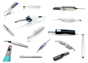
Dr. Jane, a forward-thinking clinician with an insatiable enthusiasm for digital dentistry, set out to revolutionize her practice. She hired intraoral scanners from multiple vendors, explored various brands, and partnered with several dental labs, all in pursuit of precision and efficiency. The future, she believed, was digital. But reality had other plans.
Her first battle? Edentulous scans. No matter how meticulously she positioned the scanner, the software seemed utterly confused—was it an upper jaw, a lower jaw, or a barren landscape from a sci-fi movie? When the dentures finally arrived, they fit about as well as a square peg in a round hole. One patient even jokingly asked if she had accidentally sent his scans to a shoemaker instead of a dental lab. She burst into laughter, though a part of her questioned whether she actually had.
Then came the crowns. Some fit like a dream; others were so loose they could double as castanets. “Maybe it’s a technical error,” she mused. “Or maybe my lab technician moonlights as a magician—making occlusion disappear!” She laughed at her own joke, though her patient, biting down on an unstable crown, wasn’t quite as amused.
And then, the scan bodies—oh, the scan bodies! Dr. Jane had meticulously selected the correct one chairside, yet when she reviewed the digital file, the software had chosen an entirely different entity, as if it had a mind of its own. It was like ordering a sensible sedan and receiving a monster truck instead. At one point, after hours of troubleshooting, she simply threw her hands up and exclaimed, “Maybe the scanner knows something I don’t!” Her assistant couldn’t help but chuckle.
Yet, amid the frustrations and bursts of laughter, Dr. Jane realized one thing—digital dentistry wasn’t the problem; her understanding of it was. That’s when she decided to delve deeper into learning more about them.
In the rapidly advancing domain of dentistry, the integration of cutting-edge technology has become indispensable for optimizing clinical outcomes, operational efficiency, and patient-centric care. Among the most transformative innovations in recent years is the advent of intraoral scanners, which have effectively supplanted conventional impression techniques, heralding a new era in digital dentistry. However, the selection of an appropriate intraoral scanner is not merely a matter of convenience; it is a critical decision that can profoundly influence the quality of care delivered. This discourse aims to elucidate the technical underpinnings of intraoral scanners, their clinical significance, and the potential pitfalls of suboptimal alternatives, while addressing common challenges in prosthetic fitting, such as flaring margins, loose crowns, and the limitations of scanner and software in identifying scan bodies and calculating digital separating media thickness. Furthermore, we will explore the role of each scan in enhancing the accuracy and adaptability of AI models embedded within these systems, as well as the parallels between these AI systems and large language models (LLMs) in terms of iterative learning and adaptability.
The Clinical and Technical Rationale for Intraoral Scanners
1. Enhanced Accuracy and Precision
Traditional elastomeric impression materials, while historically reliable, are inherently susceptible to dimensional inaccuracies due to polymerization shrinkage, tray distortion, and operator-dependent variables. Intraoral scanners, by contrast, employ advanced optical coherence tomography (OCT) or confocal microscopy to generate high-fidelity, three-dimensional digital models of the dentition and surrounding structures. These scanners achieve micron-level resolution, often in the range of 10-20 µm, ensuring that restorations such as crowns, bridges, and aligners exhibit exceptional marginal fit and occlusal harmony. This level of precision is particularly critical in complex restorative and prosthodontic cases, where even minor discrepancies can compromise biomechanical stability and long-term prognosis.
2. Operational Efficiency and Workflow Optimization
The adoption of intraoral scanners facilitates a paradigm shift in clinical workflow dynamics. By obviating the need for physical impressions, these devices significantly reduce chairside time, minimize material costs, and eliminate the logistical challenges associated with impression shipping and storage. Furthermore, the seamless integration of digital scans into computer-aided design/computer-aided manufacturing (CAD/CAM) systems enables rapid prototyping and in-house fabrication of restorations, thereby expediting treatment timelines and enhancing patient satisfaction.
3. Patient Comfort and Experience
Traditional impression techniques are often associated with patient discomfort, particularly in individuals with heightened gag reflexes or dental anxiety. Intraoral scanners, with their non-invasive, wand-like design, offer a markedly more comfortable experience. The elimination of impression trays and viscous materials not only enhances patient compliance but also fosters a positive perception of dental care, which is integral to patient retention and practice growth.
4. Digital Integration and Data Management
Modern intraoral scanners are not standalone devices; they are integral components of a comprehensive digital ecosystem. These scanners interface seamlessly with industry-standard CAD software platforms such as Exocad, 3Shape, Blender, and emerging third-party software like BlueSky and many others that are continuously being developed. The accuracy of intraoral scanners is increasingly dependent on these advanced software solutions, as well as AI-driven training methodologies that iteratively refine their scanning capabilities. Additionally, cloud-based data storage solutions facilitate secure, real-time sharing of digital impressions with dental laboratories, specialists, and interdisciplinary teams, thereby enhancing collaborative care and reducing the risk of data loss or miscommunication.
Common Challenges in Prosthetic Fitting and Scanner Limitations
Despite the advantages of intraoral scanners, several technical and clinical challenges persist, particularly in the context of prosthetic fitting. These issues underscore the importance of selecting high-quality scanners and software systems capable of addressing these complexities.
1. Flaring Margins and Loose Crowns
One of the most prevalent issues in prosthetic dentistry is the improper fit of crowns and bridges, often manifested as flaring margins or loose restorations. Flaring margins occur when the marginal adaptation of the restoration is inadequate, leading to microgaps that can harbor plaque, cause recurrent caries, or compromise periodontal health. Loose crowns, on the other hand, are typically the result of insufficient retention or improper occlusal design. These problems are often exacerbated by inaccuracies in the digital scan or errors in the CAD/CAM design process. High-end intraoral scanners mitigate these issues by capturing highly detailed marginal geometries and enabling precise adjustments during the design phase.
2. Inability to Identify Scan Bodies
In implant dentistry, the accurate identification of scan bodies (e.g., NP or RP) is critical for ensuring the proper fit and function of implant-supported restorations. However, some intraoral scanners and software systems struggle to automatically recognize and differentiate between various scan body designs. This limitation can lead to errors in the digital impression, resulting in ill-fitting prostheses that require costly and time-consuming remakes. Advanced scanners equipped with AI-driven algorithms and comprehensive scan body libraries can overcome this challenge by accurately identifying and processing scan body data.
3. AI Training, Transformer Models, and The Precision Factor
Let me explain this by an example. The thickness of the digital separating media—representing the cement space between the restoration and the prepared tooth surface—is a critical parameter in prosthetic design. If miscalculated, it can lead to a host of clinical problems, including over-contoured restorations, inadequate retention, or improper occlusal contacts. High-quality scanners and CAD software allow for precise control over the cement space, typically ranging from 20 to 100 µm, depending on the type of restoration and luting agent used. Moreover, recent advancements in AI, particularly with transformer models and token-based learning, have enabled more nuanced decision-making within scanning software, thereby improving accuracy over time. By leveraging AI models trained on vast datasets, intraoral scanners can iteratively enhance their precision, reducing errors related to prosthetic misfits.
The Synergy of Hardware, Software, and Expertise
The efficacy of an intraoral scanner is not solely determined by its hardware specifications; it is equally dependent on the sophistication of its accompanying software and the expertise of the dental technician operating the system. The role of AI training in these systems, coupled with software updates incorporating transformer models, further augments the accuracy and adaptability of these devices, ensuring that each scan contributes to the continuous improvement of diagnostic and prosthetic design accuracy.
IO Scanner – A Strategic Investment in Digital Dentistry
The integration of intraoral scanners into modern dental practice represents a strategic investment in the future of patient care. In an era defined by rapid technological advancement, the selection of an intraoral scanner must be guided by a thorough understanding of its technical specifications, software capabilities, and integration potential. Only through this holistic approach can the full potential of digital dentistry be realized.
Dr. Jane returned to her practice armed with newfound knowledge. This time, her scanners obeyed, her crowns fit like a dream, and the scan bodies finally made sense. One day, while reviewing a flawless digital impression, she leaned back, grinned, and said, “Well, would you look at that—I think I speak scanner now.
Author:
Dr. Syed Nabeel, BDS, D.Orth, MFD RCS (Ireland), MFDS RCPS (Glasgow), is a dedicated dental professional with a special interest in Neuromuscular Dentistry (NMD). With over two decades of experience, he has been committed to diagnosing and managing occlusal and temporomandibular joint (TMJ) disorders using a patient-centered approach that integrates the principles of neuromuscular occlusion.
Alongside his clinical work, Dr. Nabeel is deeply interested in digital dentistry and the role of artificial intelligence (AI) in dental practice. He believes that technology has the potential to enhance patient care by improving diagnostics and treatment precision. His work in this field reflects his curiosity and dedication to advancing dentistry in meaningful ways.
Dr. Nabeel also enjoys sharing knowledge and has been lecturing extensively on neuromuscular dentistry. His approach to teaching emphasizes practical insights, evidence-based methods, and a focus on patient well-being, making his sessions both engaging and valuable for fellow professionals. Additionally, he is a sought-after speaker and blogger on practice management, where he provides insights on optimizing workflow efficiency, patient engagement, and the integration of modern technology in dental practices.
Based in Mysore, India, Dr. Nabeel is the founder of Smile Maker, a practice where he strives to combine advanced diagnostic tools with personalized treatment plans to help patients with occlusal and TMJ-related concerns. With a strong commitment to continuous learning and improvement, Dr. Nabeel remains deeply invested in the evolving landscape of dentistry, always seeking ways to enhance patient outcomes with care and compassion. Reach him on dentistryunited@gmail.com

