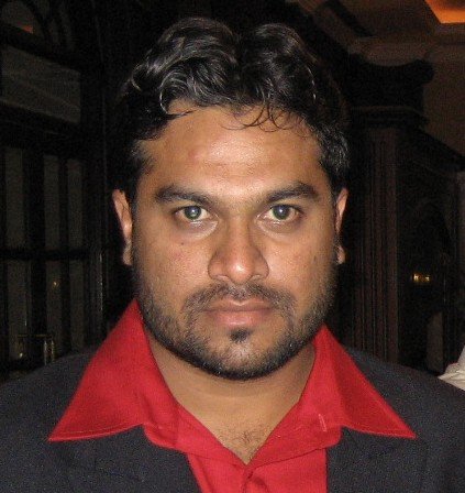
I leaned back in my chair, staring at the specs of the latest intraoral scanner on my screen. It was a minefield of jargon—CMOS sensors, AI-driven stitching, CUDA cores, and memory bandwidth. My mind drifted to an old friend, someone who had computers in his blood long before the world became obsessed with silicon and screens. Mansoor.
Back in the 90s, while most of us were struggling with floppy disks, Mansoor was knee-deep in microprocessor boards. He had seen the ZX81 of Toshiba, built and assembled 486 machines, and was the go-to guy for anything remotely technical. If anyone could demystify these scanner specs, it was him.
I picked up the phone. “Mansoor, I need your wisdom.”
He chuckled. “Still breaking teeth for a living?”
“Still putting them back together. But I need help decoding some scanner tech. These new intraoral scanners—they talk about GPUs, frame rates, and precision, but I need to know how it really works.”
Mansoor’s voice lit up, the same excitement from our younger days. “Alright, let’s start from the core. The optical system. What’s it using?”
“Mostly structured light with blue or white LED. Some older ones use lasers.”
“Ah, just like old CRT monitors struggled with color accuracy, different light sources impact what the scanner picks up.”
“Exactly! Blue LED reduces noise when scanning enamel, kind of like how an anti-glare screen works. It improves details by 10-15% compared to white light, which can cause reflection issues—think of trying to take a picture of a glass surface without glare. High-end scanners like the Medit i700 use blue LED to cut noise on reflective surfaces like enamel, achieving sub-10μm accuracy, while white light can distort occlusal morphology by 20-50μm if not optimized. In traditional impressions, it’s like using alginate that sets too fast under saliva—it doesn’t capture the true contours of a prepped margin, leading to an open margin on a crown that leaks within a year.”
Mansoor whistled. “So, it’s all about precision in data capture. What about the cameras?”
“High-end scanners use CMOS sensors, like the Sony IMX series, running at 20-30 frames per second at 2-5MP resolution. Pixel sizes below 2μm are key for capturing fine interproximal spaces. Some older ones use CCD, which has a better dynamic range for low-light areas like gingival margins but is slower.”
“That’s your bottleneck,” Mansoor said. “If your sensor isn’t fast enough, or the pixel size too large, you’ll get frame drops, which means missing data points. A CMOS with a signal-to-noise ratio above 50dB ensures clarity, but anything less introduces noise—like static on an old radio.”
“Which means distorted impressions,” I added. “A missing data point in an interproximal area can throw off the contact point of a crown by 50-100 microns, leading to either open contacts or a tight fit that requires excessive adjustments. It’s the digital equivalent of an impression tray that lifts too early, tearing the material around tight contacts—food traps form, and the patient’s back in six months with caries under the restoration.”
He laughed. “See? You’re getting it. What about processing?”
“High-end scanners use GPUs with 128-256 CUDA cores for parallel processing, like the NVIDIA Jetson in 3Shape TRIOS 5. They handle 100,000+ triangles per second for smooth 3D rendering. Low-end ones rely on weak ARM CPUs with integrated GPUs, barely managing 10,000 triangles.”
“Ah, CUDA cores—those were game-changers when NVIDIA brought them in. Parallel processing means fewer jagged edges, fewer artifacts. A weak GPU is like trying to render a 3D game on a 286 processor—it stutters, and the image falls apart.”
“Exactly. A poor GPU leads to stitching errors of 50-100μm in posterior regions. Think of it like an ill-fitted denture—small gaps accumulate, and suddenly, the occlusion is all wrong. It’s like when a polyvinyl siloxane impression sets unevenly because the tray wasn’t seated properly—distortions in the molar region throw off the bite, and the lab sends back a denture that rocks on the ridge.”
Mansoor laughed again. “Good analogy! Now, what about memory?”
“High-end scanners have 8-16GB DDR4 RAM with 256-bit memory buses, allowing 20-30GB/s data transfer. Low-end ones struggle at 5-10GB/s with 2-4GB DDR3, causing mid-scan freezing.”
“So, low RAM equals lag?”
“Exactly! When the scanner lags, it drops frames, introducing errors of 50-100 microns in posterior scans. That’s the difference between a perfect margin and a remake. It’s like taking a two-step impression where the wash material doesn’t bond to the heavy body because of a timing lag—the mismatch creates voids, and the crown comes back with an overhanging margin. Overheating compounds it too; budget scanners throttle after 5-10 minutes, dropping from 30 FPS to 15 FPS, leaving gaps in the point cloud—just like bubbles in an impression from poor mixing that ruin the fit.”
Mansoor hummed. “And calibration?”
“Premium scanners use laser interferometry for factory calibration, ensuring accuracy within 5 microns over a year. Cheaper models need frequent manual calibration with basic targets, introducing errors of 20-30 microns. Environmental factors like humidity or temperature can cause optical drift—high-end scanners compensate with temperature-stabilized optics, but budget ones falter.”
“That’s like the old CRT monitors drifting in color accuracy over time,” Mansoor mused. “So, if you don’t calibrate properly, your scans become unreliable?”
“Exactly. Imagine scanning a full arch and realizing the occlusion is off by 100 microns because of uncompensated drift. That’s a failed case waiting to happen. It’s like using an old, worn-out articulator that doesn’t hold centric relation—every adjustment in the lab is off, and the patient ends up with premature contacts that cause TMD flare-ups.”
Mansoor sighed. “So, the tech is there, but you need the right balance of hardware and software.”
“Which brings me to the software. High-end scanners use AI-driven stitching, like 3Shape’s iterative closest point algorithms with outlier rejection, reducing errors to 10 microns or less. They need only 30-40% frame overlap for seamless integration. Cheap ones use nearest-neighbor stitching, requiring 60% overlap and still introducing 50-100 micron errors.”
“That’s like early photo editing software that just guessed colors instead of properly blending them,” he said. “And AI?”
“AI helps align bite scans within 20μm using occlusal plane detection, removes soft tissue artifacts like saliva or tongue with 95% accuracy, and flags mesh inconsistencies in real-time. Without AI, manual alignment introduces 100-200 micron errors. It’s the difference between a well-seated crown and a premature contact—like an expert technician refining a wax-up versus an amateur freehanding a temporary crown. In impressions, it’s like manually correcting a distorted bite registration versus having a perfect triple-tray impression—misalignment means hours of chairside grinding.”
Mansoor let out a low whistle. “And what about color and texture? That must matter for your work.”
“Absolutely. High-end scanners use 12-bit color depth for 4096 shades, ensuring accurate shade matching with systems like Vita Classical. They apply UV mapping with sub-pixel precision to capture micro-textures like perikymata, aiding caries detection. Low-end ones with 8-bit depth and basic mapping distort colors and smooth out textures—shade mismatches happen in 15-20% of cases, and diagnostic details get lost. It’s like trying to match a shade with a poorly mixed composite impression material—you end up with a restoration that looks off under operatory lights.”
“So, you’re not just buying a scanner,” Mansoor said. “You’re buying processing power, software intelligence, and precision. Just like back in the day, when a 486 processor and a Pentium weren’t just numbers—they determined how fast and smooth everything ran.”
I smiled. “Exactly. And in dentistry, precision isn’t just about numbers—it’s about clinical success. A scanner with outdated software stagnates, while high-end ones evolve. Take the Medit i700—its 2024 update cut stitching errors from 15μm to 8μm with neural network outlier rejection. Or 3Shape TRIOS 5, which shaved a full minute off full-arch scans with GPU threading optimization. Software updates alone can turn a good scanner into a great one, saving me from the nightmares of remakes that plagued my early days with alginate.”
Mansoor chuckled. “You’ve come a long way, Nabeel. So, which one are you buying?”
“The one that ensures I don’t have to do unnecessary adjustments on every case,” I said. “Which means the best balance of optics, processing, and AI. I’m looking at the Medit i700 for its lightweight design and fast scanning, the 3Shape TRIOS 5 for its AI-powered precision, or the iTero Element 5D with NIRI technology for caries detection. But I’ll ask the right questions first—sensor specs, CUDA core count, update frequency, mesh resolution, and artifact handling.”
Mansoor laughed. “Then you’ve got your answer.”
I hung up, grateful for the trip down memory lane. The scanners had changed, but the principles of good tech hadn’t. And Mansoor, the 8MB RAM, floppy disk, 8MB CD-ROM, 1.44MB (3.5-inch) floppy disk, and 1.2MB (5.25-inch) floppy disk, along with the 486 processor-assembled computer by you, is still preserved in my office as a gift of a lifetime. You taught me back in 1994 that hardware is everything you can touch and software is everything you can see but cannot touch—and that definition still holds true today.
Notes for the Readers of This Blog
A scanner is not just a machine—it is a bridge between perception and reality. Choose wisely. The Medit i700 (lightweight, fast scanning), 3Shape TRIOS 5 (AI-powered precision), and iTero Element 5D (NIRI technology for caries detection) are not mere tools but guides in the art of restoration. When evaluating options, ask about the processor (e.g., CUDA cores, clock speed), frame rate (30+ FPS under load), and software capabilities (AI stitching, artifact removal, high-poly meshes). Low-cost scanners may falter with weak optics and static software, introducing errors of 20-50μm—errors that echo the distortions of a rushed alginate impression—while premium ones refine their craft with each update, aiming for trueness below 10μm. As with all things in life, the finer the details, the greater the outcome.
Key Hardware and Software Features with Brands:
Hardware Features:
– Blue LED Structured Light: Reduces noise on reflective surfaces like enamel, achieving sub-10μm accuracy (Medit i700, 3Shape TRIOS 5).
– CMOS Sensors (Sony IMX Series): Capture 20-30 FPS at 2-5MP resolution with pixel sizes below 2μm for fine interproximal detail (iTero Element 5D, Medit i700).
– CUDA-Core GPUs (NVIDIA Jetson): 128-256 cores handle 100,000+ triangles/sec for smooth 3D rendering (3Shape TRIOS 5).
– High-Speed DDR4 RAM (8-16GB): Supports 20-30GB/s data transfer, minimizing lag and frame drops (3Shape TRIOS 5, iTero Element 5D).
– Temperature-Stabilized Optics: Compensates for environmental drift, maintaining accuracy within 5μm (Medit i700, 3Shape TRIOS 5).
– Laser Interferometry Calibration: Factory-set for <5μm drift over 12 months (iTero Element 5D, 3Shape TRIOS 5).
Software Features:
– AI-Driven Stitching (Iterative Closest Point): Reduces stitching errors to <10μm with 30-40% frame overlap (3Shape TRIOS 5).
– Neural Network Outlier Rejection: Cuts stitching errors from 15μm to 8μm in updates (Medit i700).
– Automated Bite Alignment (Occlusal Plane Detection): Aligns bite scans within 20μm (Medit i700, 3Shape TRIOS 5).
– Artifact Removal (Soft Tissue): Removes saliva/tongue artifacts with 95% accuracy (3Shape TRIOS 5).
– High-Poly Mesh Construction (500k+ Triangles): Uses Laplacian smoothing to preserve details like incisal edges (Medit i700, 3Shape TRIOS 5).
– 12-Bit Color Depth (4096 Shades): Ensures accurate shade matching for systems like Vita Classical (iTero Element 5D, 3Shape TRIOS 5).
– UV Mapping (Sub-Pixel Precision): Captures micro-textures like perikymata for caries detection (3Shape TRIOS 5).
– NIRI Technology (Near-Infrared Imaging): Aids in caries detection alongside scanning (iTero Element 5D).
– GPU Threading Optimization: Reduces full-arch scan time by up to a minute (3Shape TRIOS 5).
Author: Dr. Syed Nabeel, BDS, D.Orth, MFD RCS (Ireland), MFDS RCPS (Glasgow), a distinguished dental professional, is the Founder and CEO of DentistryUnited.com, a pioneering platform established in 2004 to foster knowledge-sharing within the global dental community. His relentless pursuit of advancing dental education led to the creation of Dental Follicle – The E-Journal of Dentistry (ISSN 2230-9489) in 2006, a publication dedicated to scholarly discourse and contemporary advancements in the field.
As the Managing Director of Smile Maker Clinics Pvt Ltd, Dr. Nabeel oversees a growing network of dental clinics in South India, where excellence in patient care is seamlessly integrated with innovation. His clinical expertise lies in Neuromuscular Dentistry, Full-Mouth Rehabilitation, and Smile Makeovers—areas where he has transformed countless lives through meticulous, patient-centered care. Beyond clinical practice, his in-house research team is a rare testament to his commitment to evidence-based dentistry, making Smile Maker one of the few private practices globally that actively contributes to dental literature.
With over two decades of experience, Dr. Nabeel has emerged as a leading authority in occlusal dynamics and temporomandibular joint (TMJ) disorders. His holistic approach to neuromuscular occlusion places patient well-being at the forefront, ensuring precision-driven, long-lasting outcomes. Passionate about the intersection of digital dentistry and artificial intelligence (AI), he continues to explore how cutting-edge technologies can revolutionize diagnostics, treatment planning, and patient experience.
A natural educator and thought leader, Dr. Nabeel is a sought-after speaker in neuromuscular dentistry and practice management, captivating audiences with his practical insights and evidence-based methodologies. His lectures focus on workflow optimization, patient engagement strategies, and the seamless integration of modern technology in dental practices—empowering fellow professionals to elevate their standards of care.
Beyond dentistry, Dr. Nabeel’s intellectual pursuits extend to wildlife photography, travel, gardening, and creative thinking, reflecting his insatiable curiosity and deep appreciation for life’s wonders. His ability to blend science with artistry, precision with empathy, and tradition with innovation underscores his unique impact on the dental profession.

